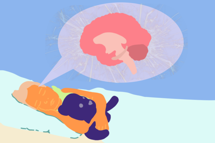
Magnetic resonance imaging used to observe hippocampal and anterior medial temporal lobe activation
By AARYA GUPTA — science@theaggie.org
UC Davis researchers from the Center of Mind and Brain recently explored how newly learned words are remembered by toddlers, according to a paper published in Current Biology.
“This is a study that examines how toddlers learn new words,” a professor of psychology at UC Davis Simona Ghetti said. “Because we are interested in both inducing some sort of learning about words and looking at the neural mechanisms that support this ability in early childhood, we created a laboratory situation where 2-year-olds could come in and experience new words associated with objects they have never seen before.”
The study followed 38 toddlers, between the ages of 25 to 32 months, as they engaged in two different tasks where they remembered newly learned words. Specifically, toddlers were taught pseudowords that were associated with “novel objects and puppets,” according to the paper.
Ghetti explains that there are two main reasons for selecting this age group.
“First, during this period, we know toddlers learn many words,” Ghetti said. “It is a period when we hypothesize that the structures — like the hippocampus, which are important for memory — would be more functional. Therefore, it is a period that is right to both examine the function of the structure and examine [its] relation with the behavior, word-learning, which is so important at this time.”
Additionally, researchers collected functional magnetic resonance imaging, or fMRI, data while these now-learned pseudowords and new pseudowords were played when the toddlers were asleep.
Hippocampal activity was recorded because it is foundational in forming relational memories, which Ghetti described to be when different aspects of an experience are put together in a representation that captures the richness of that experience.
“The hippocampus has been considered for decades a very important structure for forming and retaining associations,” Ghetti said. “[Like] what we looked at in the study, the association between [a] new word and what it refers to. Because the hippocampus is so foundational for forming relational memories, we thought that it would be particularly important to examine its role in word learning.”
Christine Wu Nordahl, a professor of psychiatry and behavioral science at UC Davis School of Medicine, contributed to the development of methods used to scan the toddlers’ brains during their sleep.
While Nordahl and her team of UC Davis researchers weren’t the first to conduct such scans, they have had a high success rate with this and past projects.
“At 88 percent, our success rate has been higher than most sites, likely because we are able to work so closely with the families,” Nordahl said via email. “Since 2006, we’ve scanned over 450 toddlers during natural sleep, many at multiple time points, allowing us to track their brain development as they grow up.”
Nordahl collaborated with UC Davis Health’s Imaging Research Center to modify scanning environments to increase comfort and decrease how intimidating magnetic resonance imaging, or MRI, machines can appear to be.
“We would plan the session around their usual bedtimes and the parents could get up on the MRI bed with their children to snuggle or read them books to help fall asleep,” Nordahl said via email. “Sometimes we would have children fall asleep in the car on the way over to the Imaging Center and simply carry them into the scanner and place them on the bed directly, already asleep. We let the parents stay in the room with their child the whole time and we would also stay in the room watching the child during the scan in case they woke up. If they did wake up, we would be able to stop everything right away.”
This process was critical for their study.
“Scanning during natural sleep allows us to examine brain structure and function during this critical window of development,” Nordahl said via email. “If a child this age needed an MRI scan for clinical purposes, they would usually undergo general anesthesia in order to remain still enough to get a picture of their brain. But for research, general anesthesia is not allowable because of the increased risk.”
Graduate student Lindsey Mooney was one of the UC Davis students involved in this research.
“I […] really enjoy being a part of work that is actively pursuing answers to unanswered questions about child development,” Mooney said.
Mooney said her responsibilities included project design, data collection and data analysis.
“Getting small children to fall asleep and stay asleep in the scanner is challenging, but that makes the data and results so much more exciting and rewarding,” Mooney said.
Overall, this research examines the brain mechanisms associated with memory, particularly in early stages.
“We can show that even very young children have quite sophisticated memory processes that they can put into service to form memories that are quite complex like words or events about their lives.”
Ghetti expands that several auxiliary questions can be asked from this research.
“We can now ask even more precise questions about the nature and retention of memory in young children,” Ghetti said. “We might have some new insight in how children form vocabulary. Communicating and speaking is so foundational for children’s capacity to interact with loved ones, the world and to learn. We can contribute to understanding how that happens.”
Written by: Aarya Gupta — science@theaggie.org


