

UC Davis Immunology researchers engineer a bone marrow to research early osteosarcoma
Through macrophages and bone cancer cells, researchers hope that the new 3D model will help them better understand osteosarcoma growth
By MONICA MANMADKAR — science@theaggie.org
UC Davis researchers employ tissue engineering to study the interaction of macrophages, a special type of immune cell involved in the detection of foreign bacteria and tumors. There have been many studies that linked macrophages to osteosarcoma progression. In this study, researchers delved into how studying these macrophages could be a potential opportunity to develop immunotherapies.
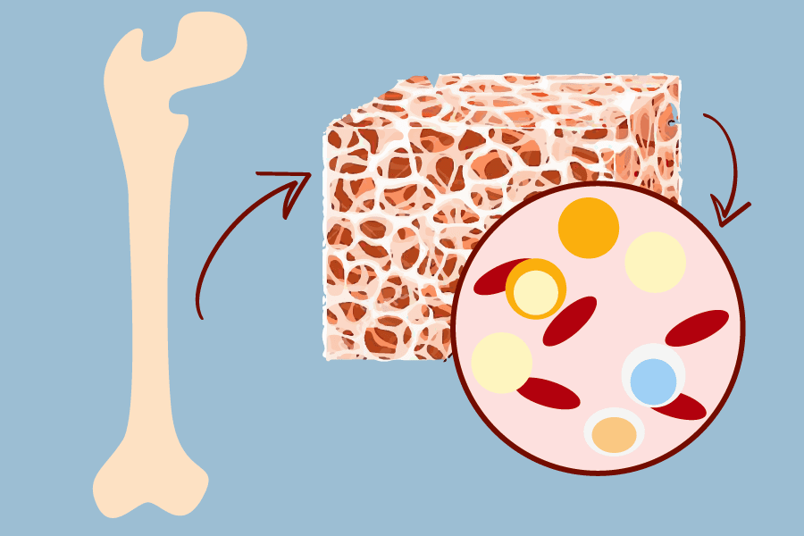
Given this understanding, the Leach Laboratory at the UC Davis Department of Orthopaedic Surgery created an artificial bone marrow model that could help them better understand osteosarcoma, a common type of bone cancer in children.
Osteosarcoma begins to develop in the bone marrow but can spread to other bones around the body, especially around the knees and upper parts of the arms. With its treatment barely making progress, less than 25% of people with this cancer survive five years post-diagnosis. This cancer grows in a complex bone marrow environment, making it harder to study. Current models do not account for its key features and 3D structure. Hence, the researchers at the Leach Lab created an engineered bone marrow (eBM) to mirror the native bone marrow and study the tumor’s progression.
“The eBM provides a 3D environment as opposed to 2D, which can greatly affect cell behavior, and since it can be maintained in vitro, it gives us a culturable way to control microenvironmental factors like oxygen tension,” Katherine Griffin, a dual doctoral of veterinary medicine and immunology candidate and a researcher at the Leach Lab, said. “There aren’t many models out there that are capable of this. It’s very novel to approach a cancer immunology question with a tissue engineering approach.”
The lab will use the eBM to study osteosarcoma cells with and without macrophages added. Macrophages are extremely important in creating the tumor’s environment and their addition can be related to disease development. Using dyes, the researchers will record the movement of the cells over time.
“We are combining immune cells which would be commonly interacting with the tissues and adding macrophages that have different characteristics,” Dr. Kent Leach, a professor of orthopedic surgery and biomedical engineering at UC Davis, said. “Adding these different types of immune cells may be contributing to tumor growth, which is a new area in the fight against cancer.”
Leach hopes to create a combination of cancer biology, tissue engineering, immunology and patient care through his laboratory’s research and future work.
Bringing immunology knowledge to the table, Griffin works as the main graduate student on the project although this is a combined effort between the faculty collaborators, Dr. Kent Leach, Dr. Steven Thorpe, Dr. Lor Randall and Dr. William Culp.
Osteosarcoma is rare enough that clinical trials and other more common methods for cancer research are less effective at efficiently figuring out its biology.
“The current model is only for mouse cancer cells hence [we] would like the eBM to translate to human and canine samples so we can really capture those natural populations and better understand patterns in osteosarcoma tumor progression,” Griffin said about their future research goals.
Written by: Monica Manmadkar — science@theaggie.org