

Researchers, radiation expert, office of research discuss implications of total-body scanner.
Researchers at UC Davis recently received a $15.5 million grant to build the world’s first total-body positron emission tomography (PET) scanner, an advancement in medical technology that researchers predict will revolutionize the health field.
After 10 years and 14 grant proposals, researchers were able to lay the groundwork for their “explorer project.”
“I feel elation and tremendous excitement when I think of the things we can do with this scanner,” said Ramsey Badawi, lead researcher and associate professor in the department of radiology at the UC Davis Medical Center. “I think we’re really going to change the way that we approach human imaging science, medicine and healthcare. There have been PET scanners before, but not great, big, long ones like this that [will be] so incredibly sensitive and capture the entire body in one go. That’s never been done before. This isn’t an incremental change — we’re going to make a really, really big change.”
PET scanners are already used clinically; however, they do not encompass the whole body. This leads to blind, or off-the-chart areas, that make it difficult to view how drugs affect the entire body or map the spread of disease.
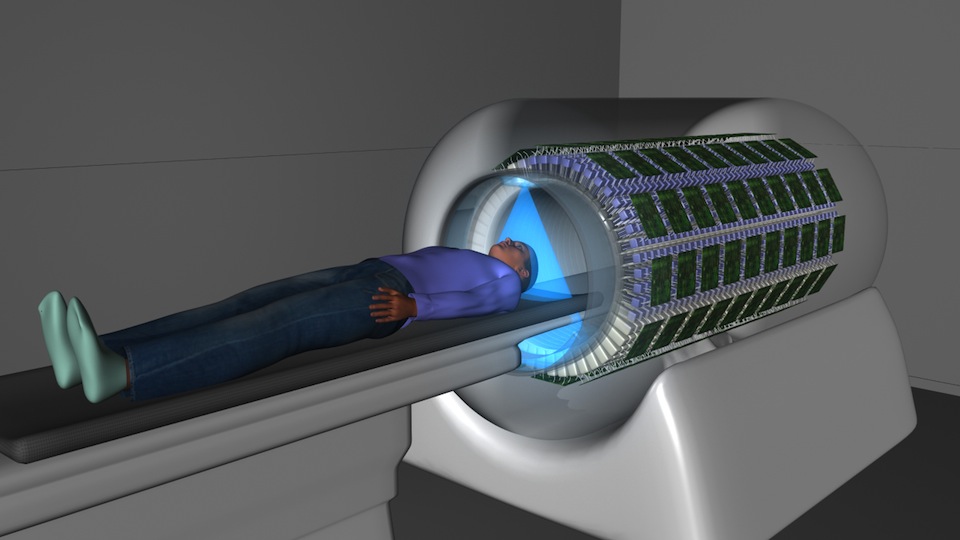
“The ring of detectors in the typical PET scanner is maybe only about 20 centimeters big, and people are typically quite a lot bigger than that,” Badawi said. “Most of the person is actually sticking out of the end of the scanner, [and] it then looks like the marker is going nowhere. If you want to image the whole person, you have to step the person through the scanner which would take a bit of time. Now with a two-meter scanner, you can fit a whole person right through. This leads to a huge increase in the amount of radiation signal we can pick out.”
While magnetic resonance imaging (MRI) and computed axial tomography (CAT) scans look at body structure, PET scans look at the function cells are carrying out. In PET scans, a small amount of a radioactively-labeled probe is injected into the body. This chemical compound is designed to find a specific target in the body. This radioactively-labeled material emits radiation, which the PET scanner detects and measures. Doctors can track where the chemical compounds accumulate to determine whether the cells are alive and dividing, dividing excessively or dying out. A mathematical algorithm is used to create 3-dimensional maps of the body.
“By changing the chemical compound, we can look at different aspects of the biology going on,” said Simon Cherry, lead researcher of the study, distinguished professor in the department of biomedical engineering and director at the Center For Molecular and Genomic Imaging. “One of the things it’s used to look at is glucose metabolism. Glucose is a sugar the cells of the body use to produce energy. In many diseases, the metabolism of glucose is altered, so it goes up or down. It goes up in cancer. [I]n some neurological disorders, like Alzheimer’s disease, cells are dying off so they have a lower use of the radioactive glucose.”
Like MRI and CAT scans, the PET scan in current use only shows one part of the body at a time. Having a total-body scanner would allow doctors to see all the organs and tissues of the body at the same time.
“This is important because a number of diseases involve the whole body,” Cherry said. “Cancer is one obvious example because when cancer spreads, you don’t know where exactly it’s going to spread. Infection and inflammation can also occur in many parts of the body.”
With the whole patient enclosed in the scan, researchers can take “movies” of how radioactively-labeled material moves around the body. This is particularly important in drug trials. By attaching radioactive material to small samples of drugs, researchers can track a drug’s movement through a patient’s body and determine if the drug will go to other organs and cause any toxic side effects.
“This is quite a big deal because it costs billions of dollars to bring a drug to the market and a lot of the drugs fail when they’re doing wide-scale testing, which is very expensive,” Badawi said. “If we can weed out the bad drugs that are going to fail earlier, we can save a lot of money. We’ve got to get really good at making drugs cheaply and this kind of technology can really help.”
This opens up many possibilities in the healthcare field. Environmental toxins are another area of concern that require more research. By introducing toxins to the body at very small, non-harmful levels, researchers can track their movements and effects in the body.
Using a total-body scanner, as opposed to the current scanner, can also significantly reduce radiation levels.
“We can do scans for 40 times less radiation with the new scanner,” Cherry said. “That’s about the same as the radiation dose that you get flying at high altitude in a plane from San Francisco to London and back again. The radiation dose with the current scanner is 40 times the dose you’d get on a transatlantic flight. One of the ideas of this new scanner is that the radiation doses are very low and in the range of things we experience in everyday activities.”
With the current PET scanner, the radiation dose is approximately equal to the amount of radiation people in the U.S. receive in two years from just living with radioactive materials in the soil and from outer space according to Jerrold Bushberg, clinical professor of radiology, radiation and oncology at the UC Davis Medical School.
“The normal risk of developing cancer in the U.S. is 44 percent,” Bushberg said. “The current PET scan can increase that by 0.1 percent to 0.5 percent depending on the gender of the patient and the age at which they were scanned. This is a very low dose, and it takes pretty high doses to increase cancer risks substantially.”
While the radiation risk associated with current PET scans isn’t high, according to Bushberg, the risk does increase for lower-age patients.
“Cancer has a time latency in development,” Bushberg said. “That’s about 20 years. Old individuals would likely succumb to other disease processes first, so the benefits of a PET scan dramatically outweigh the risks. Even for younger patients, if they do need a medical imaging procedure that is well justified and will play a role in their care, the radiation risk is secondary.”
One of the benefits of the lower doses of radiation with the new scanner has to do with giving children a less risky health method.
“PET scanning in adults has produced some pretty important results and many important advances in health care,” Badawi said. “It’s less available to kids because kids are more radiation-sensitive. So we don’t do the kind of research we do on adults on kids. If you’ve got a scanner with a much less radiation dose, now we can do science that can lead to cures that the adults would have to take drugs for.”
Because radioactive material has a half-life, meaning it decreases in quantity over time, materials with shorter half-lives can only be used for a short amount of time before they’re gone. A regular PET scanner works for approximately three half-lives, allowing for about six hours of imaging. The new PET scanner would work for eight half-lives, allowing imaging for 16 to 18 hours.
“Some agents that last for three days, we should be able to trap them in the body for a month,” Badawi said. “We can look at disease over a long period of time without really putting them at any radiation risk. […] If you get an ACL injury, you’ve got a pretty good chance of getting arthritis in 10 years following that. We can start to do PET scans on patients to look at arthritis development, or how that injury progresses.”
According to Cherry, the full-body scanner allows researchers to take the image in one-fortieth of the full time (or 30 seconds) while still using the full radiation and achieving high quality images.
Current estimates predict building of the scanner will be finished in two to three years. After that begins the long process of research and collecting data. If the project is successful, researchers hope the scanners will reach other major research institutions on a global scale.
“The goal is to show the impact of this technology on medicine and medical care,” Cherry said. “Building the scanner is really just the first part. What matters is how we’re going to use it. The goal is to get into doing studies in humans as quickly as possible, doing better cancer imaging, tracking an infection, helping to develop new drugs. We see this as being mostly research in the beginning. Before it can go to hospitals, we have to prove the value it can bring to modern medicine.”
While Badawi and Cherry are principal investigators in this project, the research also involved teams from a multitude of departments and was a joint effort with the University of Pennsylvania and the Lawrence Berkeley National Lab. Cherry and Badawi credit much of their effort to the Research Investment in Science and Engineering (RISE) program at UC Davis.
“[This] project really epitomizes our hopes for the RISE program,” said Paul Dodd, associate vice chancellor in interdisciplinary research and strategic initiatives at Office of Research. ”This project stood out as perfectly fulfilling our desire to stimulate high-risk, high-reward interdisciplinary research with a potential to have significant societal benefit. It was one of the outstanding concepts from our faculty selected as one of 13 proposals funded from 119 applications after a rigorous review process.”
While the process will be long and a great deal of responsibility, Badawi and Cherry both look forward to exploring the applications of their project and leaving immensely positive impacts within the medical field.