
Innovative technology uses plastic to create exact replica of complex heart
The UC Davis Medical Center recently utilized 3-D printing technology to construct a three-dimensional model of a patient’s heart to assist surgeons. The 3-D heart follows other plastic printing breakthroughs, such as prosthetic ears and limbs.
The technology is the result of a complicated pediatric heart surgery case from January, where a 19-month-old girl was born with a complex constellation of arteries coming off her heart and only 1.25 ventricles rather than the usual two.
A group of UC Davis specialists consulted with biomedical engineers from Translating Engineering Advances to Medicine (TEAM), a university engineering laboratory that specializes in medical prototyping. Together, they were able to construct an exact 3-D-printed replica of the patient’s heart.
TEAM’s state-of-the-art facilities allowed the heart center to rapidly print the model in a rubber-like proprietary plastic mix that mimics the heart’s pliability. By using previously obtained MRI and CT scans, the biomedical engineers were able to form an image of the heart and send it to the 3-D printers.
“Scans are given to us, and we have to take [those] and we have to define boundaries of what is printed or what is blank space,” said Jerry Hu, TEAM’s principal development engineer for biomedical engineering.
Hu recognized that the 3-D model provided unparalleled detail that cannot otherwise be achieved with only scans.

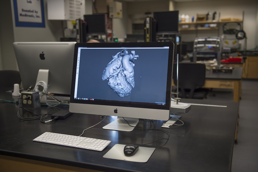
“When you see the scans on the monitor, they’re in sections and are flat images. It’s quite different than holding it in your hand,” Hu said. “Playing with [the model] is different than looking at 2-D images or 3-D models on a computer.”
The detail of the 3-D heart model helped pediatric heart surgeon Gary Raff decide on the safest and most effective surgical procedure. Raff successfully performed an aortic translocation and removed the obstruction under the pulmonic valves and aortic valves. The physical model provided Raff the intricate details needed to fully assess the condition of the patient.
“This is a tremendous resource and a unique one, something that could only have happened with the collaboration between the Davis campus and the medical center,” Raff said in a press release. “It can really help save lives.”
The technology of 3-D printing has been utilized previously at the UC Davis Medical Center. In a previous cardiothoracic case, 3-D modeling helped surgeons analyze a tumor surrounding a patient’s heart. Surgeons were able to assess the complexity of the tumor, ultimately deciding that surgery would be too complicated to perform. They then were able to proceed with a treatment plan that was less risky to the patient.
This technology has yet to become as common as MRI scans or CT scans, and has only just recently become widespread within the surgical community. Issues of cost and access make this technology underutilized, but Steven Lucero, mechanical engineer and TEAM facility manager, hopes this will change within the next 10 to 15 years and allow facilities like TEAM to become more common.
“Technology like this is still fairly expensive and rare. There are only a few facilities like ours,” Lucero said.
Written by: Lindsay Floyd – campus@theaggie.org