
New research improves noninvasive brain imaging
What might a stroke patient, a drowning person, and someone with severe traumatic brain injury have in common? If the stroke results from a blood clot in the brain, then all three might experience changes in their cerebral circulation. Blood flow in the brain is an important indicator of its health; proper blood pressure and circulation means enough oxygen and glucose are reaching the cells, and they require a lot of circulation to stay healthy.
Traditional methods for measuring cerebral blood flow and pressure include MRI and probes. However, they come with their own caveats. Metal and electronic items should not be taken inside MRI machines, including implants, prostheses, and pacemakers. This procedure is thus limited to people who don’t have those items, and is also relatively expensive, making conditions where multiple scans are necessary costly. Probes are invasive, as they must be inserted into the head to sense pressure.
One alternative is diffuse correlation spectroscopy, where certain wavelengths of light are able to penetrate into the brain and create an image. This procedure is noninvasive and can be used at the bedside, allowing for continuous monitoring. However, the spatial resolution isn’t very good compared to an MRI, and the materials needed to make it so are very expensive. Currently, DCS is not commonly used in medical practice.
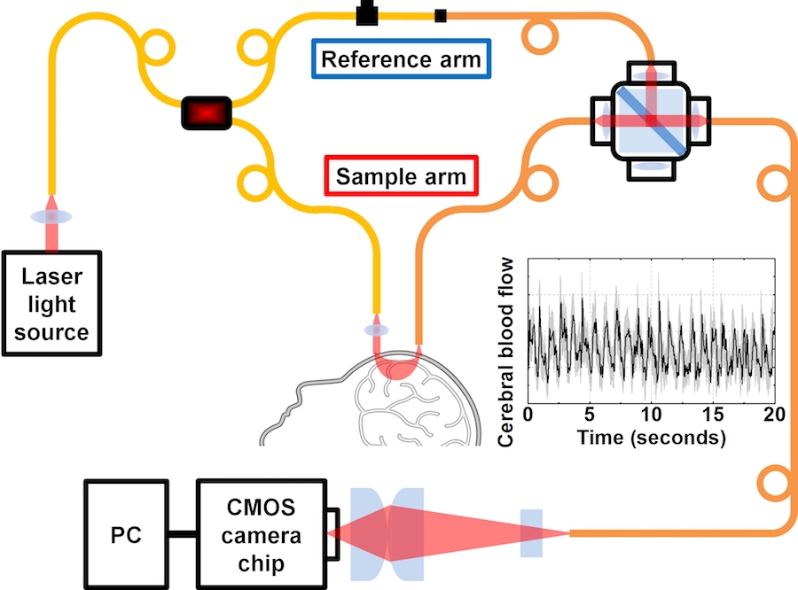
New research from a biomedical engineering lab at UC Davis may help change that. Wenjun Zhou, a postdoctoral fellow working in Vivek Srinivasan’s biophotonics lab, and his colleagues have developed an improved light-based method for measuring cerebral blood flow they call interferometric diffusing wave spectroscopy, or iDWS.
“The previous methods required very expensive detectors,” said Srinivasan, an associate professor of biomedical engineering and ophthalmology at UC Davis. “We looked at that method, and while it worked, the problem was that you couldn’t have many detectors detect the light, because it would be too expensive; the technology is somewhat difficult to use; and it’s sensitive to ambient light, or environmental light, which makes it difficult to use if you want to monitor blood flow in a hospital environment where the lights are very bright. So we tried to improve the detector, and we came up with this idea, which is really an combination of two ideas. The first idea is that if you think of what the cheapest, most widely used light detection device out there, it’s got to be the CMOS camera.”
The CMOS camera is the type of camera found in smartphones, which uses a sensor chip to record images. It is easily integrated into other technology, but its resolution isn’t as fine, so it wouldn’t be able to pick up the weak light used for DCS. To resolve this problem, the researchers changed the strength of the light field without strengthening the source.
“Measuring blood flow centimeters below the surface has required expensive approaches to count the small numbers of light particles, or photons, that make it back,” Zhou said in an email interview. ”Boosting the light power on the scalp is not possible due to concerns about heating. Instead we use an interferometric approach to boost the weak sample light field with a strong reference field, creating intensity fluctuations that are strong enough to be measured by a inexpensive CMOS camera.”
The combination of multimode fiber interferometry and CMOS cameras is a promising one; not only does it cut much of the cost, but it also has highly sensitive and parallel detection capabilities for faster and deeper imaging. It also has the potential to become better as CMOS camera quality continues to improve at a fast rate. The lab is currently collaborating with doctors at the UC Davis Medical Center to further develop this technology, including Lara Zimmerman, an assistant professor in the department of neurological surgery and neurology at UC Davis and co-director of the Neurocritical Care Service.
“When the brain is injured, it undergoes a period of swelling and the pressure inside the skull (intracranial pressure) increases,” Zimmerman said in an email interview. “This is a very dangerous problem which can lead to additional brain injury due to lack of blood flow and oxygen delivery to the brain. Currently we are able to monitor intracranial pressure with invasive probes, but ideally want to develop a technique to monitor cerebral blood flow non-invasively, which is the goal of this collaboration.”
Written by: Kira Burnett — science@theaggie.org