

Scans with specially designed PET system for horses successfully executed by Veterinary Medical Teaching Hospital
UC Davis VMTH veterinarians, professors created PET scanning system to identify equine lesions
The UC Davis Veterinary Medical Teaching Hospital (VMTH) has successfully administered scans for horses using their new equine-specific PET system. The machine allows for the 3D scanning of horse limbs to detect lesions and injuries that other technologies cannot identify, according to Mathieu Spriet, an associate professor of diagnostic imaging at the UC Davis School of Veterinary Medicine.
On Oct. 8, VMTH had the first safety demonstration, illustrating that a horse can safely step out of the machine if they move. So far, four horses have been scanned with the machine, and another set of horses will be scanned this week.
PET (positron emission tomography) provides a 3D representation of tissue and is useful for detecting sites of high metabolism associated with injury and healing in bone, according to an email from Susan Stover, a professor in the department of surgical and radiological sciences in the School of Veterinary Medicine. Radioactive material is injected into an area, creating bright spots on the scanned images to show the injuries.
UC Davis specifically developed this new machine to be used with racehorses. In racing, the primary injuries involved are in the sesamoid joint, which is in the horse’s ankle, or fetlock. “Breakdown,” which occurs when horses become very injured in their fetlocks through repeated racing, makes up about 50% of the fatal injuries in racehorses. It is very difficult for the horse to survive these kinds of injuries, said Rick Arthur, the equine medical director at the School of Veterinary Medicine.
“There’s a lot of catastrophic injury with horses breaking down in the fetlock,” Spriet said. “That is why the focus was on the fetlock in the first study.”
PET scanning can identify preexisting lesions and horses that are predisposed to having these lesions. Certain types of lesions are not visible through other diagnostic techniques, according to Arthur. The goal is to avoid racing horses with these injuries to keep them sound, which is a term to describe a healthy horse.
“When we measured like 20 racehorse fetlocks, we saw a bunch of lighting up in the sesamoid bones in the fetlock,” Spriet said. “That is why it is very interesting to use it to better understand what is happening in these cases so we can change what we are doing before the horses break down.”
Since the machine is a round structure, it collects 3D information by scanning all the way around the limb. Other scanners such as ultrasounds, scintigraphy and radiographs only take 2D images, so they cannot identify all injuries, Spriet said.
“It may look normal in other scanners, but PET uses radioactive tracers,” Spriet said. “With molecular imaging, you can see some changes at the microscopic level, which is really important for us. Once you have a big hole in the bone, it is hard to repair, but if you can detect that something is happening before a big hole develops, that helps.”
The idea for using the technology for horses was developed about six years ago. A bioengineer recommended to use his newly developed PET scanner for horses, as he was writing a grant and was trying to show as many applications for the scanner as possible. Although that system did not end up working for horses, it gave Spriet the idea to use PET with horses. PET is usually used in oncology for detecting cancerous tumors in humans and other animals, so it was never really on Spriet’s radar.
“That was the first time the idea came, so we are very grateful for [the bioengineer],” Spriet said.
Four years ago, VMTH got its first PET scanner. It had been developed for human heads, but by placing it on a cart, a horse’s limb could fit in it. Horses had to be anesthetized and laid down in order to use the machine, since the scanner had to go all the way around the horse’s limb, Spriet said..
Horses should not be put under general anesthesia very often, however, because it is a dangerous and complex process, according to Spriet. With this machine, it would not be safe to use with a standing horse, since the horse’s limb could not get out of the cylinder if they moved. Therefore, they wanted to develop a system that they could use with a standing horse that would detach if a horse moved.
“We did not want to bring [the machine] further up the limb because even if the horse is sedated, at some point they are going to want to move,” Spriet said. “We needed something that will open up.”
With a standing unit, the procedure is easier, faster, cheaper and less risky to administer, so clients will be more willing to use this type of diagnostic imaging, according to Arthur.
Last January, Spriet’s team created the first standing machine, however it was only able to scan the hoof. The machine involved the horse standing in a shallow hole, since it would not be safe for the horse’s whole leg to be encased in the machine, according to Spriet.
Last spring, the team received funding to design and build a machine that disengages when a horse move. The arms open up so the horse can get out of the machine if they move.
“The detectors are the same but the scanner itself is divided into three parts, and so it goes up and down on the limb to the foot, the big safety concept we need the horse to be able to step out of that if he gets stressed,” Spriet said.
Now, the VMTH has two PET machines, one where horses lay down under general anesthesia and the new one where horses can stand up. With the standing machine, the images are a little more blurry since the horse has more motion, but the lesions can still be seen very clearly.
“What we can find under anesthesia, we can also find in the machine without,” Spriet said. “We are still exploring what is the best protocol and way to scan to be the most efficient. We are pretty excited about what we have demonstrated.”
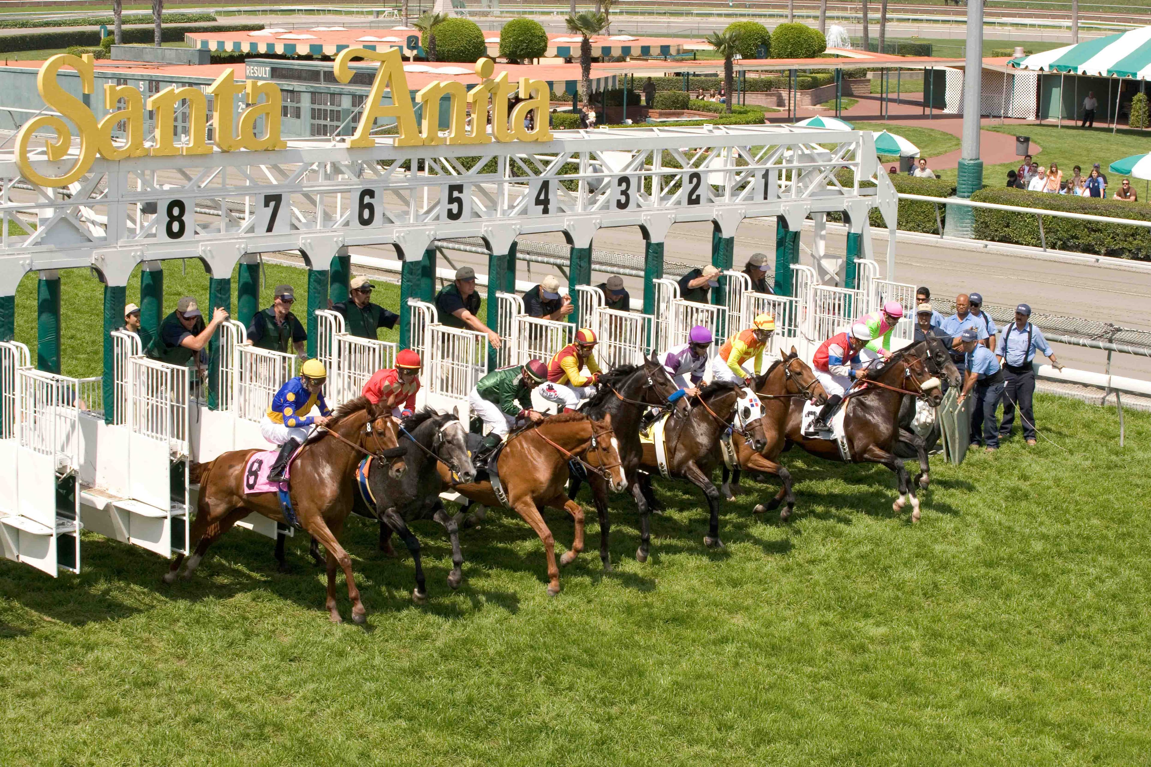
The standing PET machine will not stay in Davis for long. Since the funding for the machine came largely from the Grayson Jockey Club and the Santa Anita race track, the machine will move down to Southern California. It will be transported to Santa Anita either in the last week of November or the beginning of December.
The next machine they want to develop is a hybrid of the two previously developed systems. Spriet said they want a system that opens up so standing horses can use it but that also has the ability to rotate 90 degrees so anesthetized horses can use it as well. This machine will stay at UC Davis to be used for clients.
“Typically we will do radiographs and ultrasounds and then MRI,” Spriet said. “This will go instead of MRI or when MRI does not fully answer what is going on. These are more advanced cases. It will help us figure out what kinds of lesions are present, see what is happening and whether the racehorse needs some more rest or needs to change something in the training to reduce stress.”
Longmile Veterinary Imaging builds the PET machines that UC Davis uses. Not many places offer the technology yet, according to David Beylin, the executive director of Longmile Veterinary Imaging. In the next year Beylin said there will be more sites offering the technology.
This technology will be implemented in other contexts than racing and will be especially beneficial to other horse sports such as jumping, dressage and western events, according to Stover.
“It is not going to be the magic thing that will solve all the problems, but it is one tool to really help understand the problem better,” Spriet said. “Ultimately, it will prevent the breakdown of horses and keep horses healthier and racing longer.”
Written by: Margo Rosenbaum — science@theaggie.org