This emerging technology will improve medical diagnoses and anatomical knowledge
By KATIE HELLMAN — science@theaggie.org
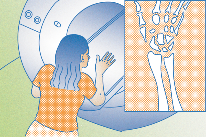
A recent study published in the British Journal of Radiology has shown that a new magnetic resonance imaging (MRI) system can capture the live motion of wrists. These new systems can lead to a better understanding of the anatomy of human wrists and can advance the future of diagnostic technology.
“A detailed understanding of the functional kinematics of wrist tissues necessary to carry out activities of daily living is essential to effectively diagnose and treat wrist dysfunction,” the study reads. “Real-time MRI of the moving wrist is feasible with high-performance 0.55T (Tesla) and may improve the evaluation of dynamic dysfunction of the wrist.”
Abhijit Chaudhari, a professor in the department of radiology and interim director of the UC Davis Imaging Research Center, commented on the value of this new advancement in an interview with UC Davis Health.
“Moving images give us a new tool to diagnose wrist dysfunction, either during motion or when there is load on the joint,” Chaudhari said. “The wrist is highly complex, so the ability to visualize motion will have an enormous impact.”
Traditional still MRI images usually provide highly accurate information regarding the wrist’s internal structures.
“Magnetic resonance imaging (MRI) has high spatial and contrast resolution, and can [characterize] bone and soft tissue without using [ionizing] radiation, making it an ideal imaging modality to assess pathologic conditions affecting joints,” a study published in PubMed states.
However, despite their reliability, current MRIs have a major fallback: still images can make it difficult to detect dynamic instability, which refers to unstable joints that are only visible during movement. Assessing wrist function while the wrist is in motion is the key to diagnosing certain injuries and administering subsequent treatment.
Along with Chaudhari, Robert Szabo, a professor of orthopedic surgery at UC Davis, and Robert Boutin, a musculoskeletal radiologist and professor of radiology at Stanford University, have spent over 10 years developing three Tesla (3T) machines to create live scans of the body. Despite the development of this fascinating technology, using a high-field strength magnet created artifacts in images that made it hard to obtain a clear reading of the MRI scans.
To solve this problem, Chaudhari, Szabo and Boutin began working with Krishna Natak, director of Dynamic Imaging Science Center at the University of Southern California. They received a grant to create a 0.55T MRI system that can develop moving images at 78 frames per second. This allows for precise dynamic imaging in a matter of seconds without artifacts obstructing the ability to interpret radiology reports.
“The dynamic pictures, along with standard, still MRI scans, show us specific wrist anatomy that had never been evaluated to this degree before,” Szabo said during an interview with UC Davis Health. “This has tremendous relevance to evaluate injuries and conduct further research into how the wrist functions.”
Written by: Katie Hellman — science@theaggie.org


