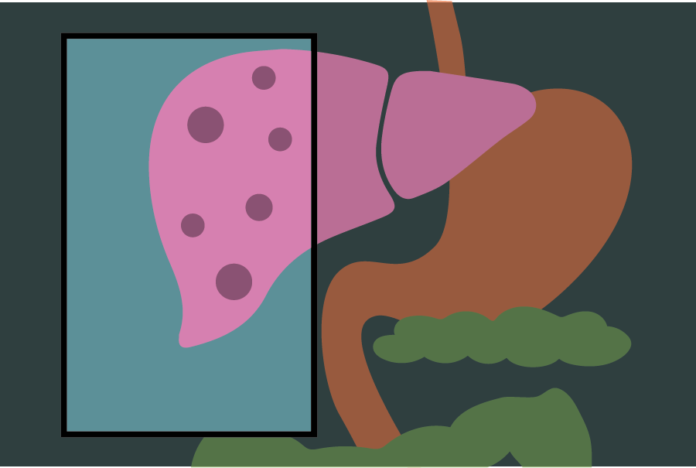
PET imaging tool provides a non-invasive alternative to liver biopsies, the current standard of detection for liver inflammation
By BRANDON NGUYEN — science@theaggie.org
According to the American Liver Foundation, about one-third of the U.S. population is diagnosed with non-alcoholic fatty liver disease (NAFLD). A severe form of this liver disease is non-alcoholic steatohepatitis (NASH), which forms as a result of metabolic fat buildup in the liver, leading to inflammation of the organ and potential scarring, or fibrosis. If not treated, the inflammation can progress to liver cirrhosis and even increased risk of liver cancer.
Dr. Souvik Sarkar, a gastrointestinal and liver specialist at the UC Davis Medical Center, described what non-alcoholic fatty liver disease entails and why it demands so much attention.
“Fatty liver disease has three parameters which define the disease, one of which is a fat, the start of the disease,” Sarkar said. “It is becoming a very fast growing problem in our country today, being driven by obesity and diabetes. The only way you can diagnose NASH at this time, unfortunately, is with a liver biopsy. There are new tools coming out to determine NASH, but they’re still in research.”
Fortunately, UC Davis Health researchers and clinicians have introduced a non-invasive alternative to the invasive liver biopsy to detect fatty liver disease — a positron emission tomography (PET) imaging tool. Dr. Ramsey Badawi, vice chair for research in the Department of Radiology and the co-director of the EXPLORER molecular imaging center at the UC Davis Medical Center, discussed why the new PET imaging tool for liver disease is so significant.
“With this new technique of processing the data using the PET scan, clinicians can easily detect an inflamed liver without having to do a biopsy,” Badawi said. “That’s a huge deal because it’s like 100 million people we’re talking about. You can’t do biopsies on all these people. That is why this non-invasive way of detecting fatty liver disease is so significant.”
Dr. Guobao Wang, an associate professor of radiology at the UC Davis Medical Center and a co-lead of the project alongside Dr. Sarkar, echoed Badawi’s optimism toward this first-of-its-kind non-invasive method to detect fatty liver disease.
“It is exciting to see that our methodology can fill the gap in clinical imaging of nonalcoholic fatty liver disease,” Wang said. “This collaboration has been extremely fruitful, as our team has been working diligently to develop this technique.”
With the state-of-the-art EXPLORER total body PET scan machine located at the UC Davis Medical Center, both Sarkar and Wang are looking to incorporate their computational PET imaging tool into the EXPLORER machine to reduce radioactive dosage on tracers while still being able to effectively assess liver inflammation and fibrosis early on in the treatment process.
“You have a patient which you see, and we send the patient in for a scan in the EXPLORER so eight years down the road we can actually be able to predict, not only the disease that is happening in the liver, but also the disease and the potential for future disease in the heart and the brain and the kidney all at the same time,” Sarkar said. “So you can imagine what a big benefit that will be for the patient being able to plan their health with more personalized care.”
Written by: Brandon Nguyen — science@theaggie.org


