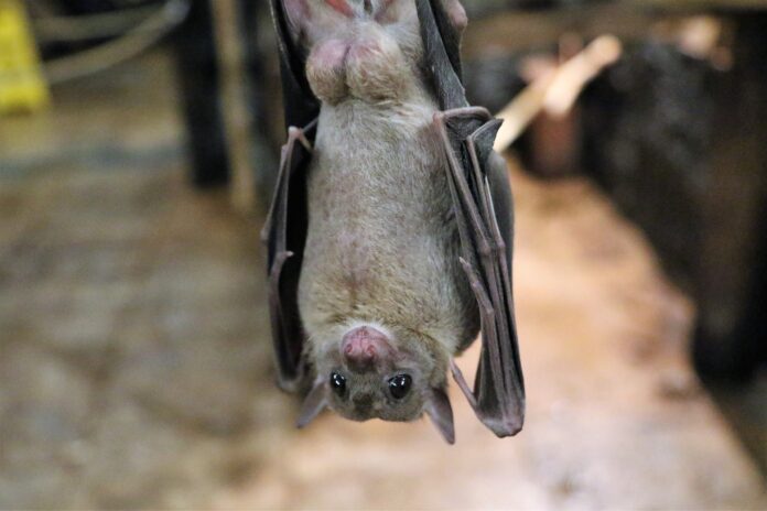
New research by Dr. Andrew Halley and the Krubitzer Lab at UC Davis details how bat brains are highly specialized for echolocation and flight
By MARGO ROSENBAUM — science@theaggie.org
People often wonder how the mammals we see swinging through trees, swimming in the ocean or flying over our heads relate to us. We ponder how millions of years of evolution resulted in so many mammals of varying intelligence and abilities.
If only we could look right into their brains.
Dr. Andrew Halley, a postdoctoral researcher in the Krubitzer Lab at UC Davis, did just that. With the help of fellow researchers at UC Davis, Simon Fraser University and UC Berkeley, Halley performed brain surgeries on anesthetized bats to better understand the motor cortex — the region of the brain controlling voluntary movement across the body.
Publishing the results on May 25 in the journal Current Biology, Halley and the other researchers discovered that bats’ brains are highly specialized for two unique aspects of their biology: echolocation and self-propelled flight.
To make this discovery, Halley, the lead author of the paper, and his colleagues mapped the brain regions controlling movements in these fruit bats, focusing on areas dedicated to echolocation and flight.
Bats represent a quarter of all living mammalian species, but until only recently, much of their brains and evolution remained a mystery. Halley and his fellow researchers sought to change that.
Before this study, a bat species’ full motor cortex had never been mapped. This achievement now allows researchers to understand the part of the brain involved in the planning, control and execution of voluntary movements.
Paths to studying evolution
Fascinated by evolutionary questions, Halley studies evolutionary neurobiology and comparative neuroscience. Originally from Philadelphia, he majored in psychology and worked in a genetics laboratory as an undergraduate at Pennsylvania State University. Halley said he grew up more interested in the humanities but always held a fascination for psychology.
Biology piqued his interest when he started taking biology classes in college, especially after learning about evolutionary theory. With the questions he started asking, he realized he needed to learn more about neuroscience to answer them and wanted to study brain evolution.
Halley completed his Ph.D. at UC Berkeley in 2016, after studying biological anthropology and working on a project tangentially related to neuroscience, in which he studied differences in embryonic development across species.
This fascination for evolution and neuroscience brought him to the Krubitzer Lab at the UC Davis Center for Neuroscience as a postdoctoral researcher.
“The Krubitzer Lab was sort of a natural fit; [Dr. Krubitzer] is one of the preeminent brain evolution researchers that’s around,” Halley said.
Dr. Mackenzie Englund, a former graduate student in the lab and co-author of the paper, shares Halley’s appreciation for evolution and sensory systems, which he said “are this medium through which we interact with the world.” Englund came to UC Davis for his Ph.D. to research similar questions.
“Evolution was always just one of those things that made me feel really close to the world,” Englund said.
Straying from the study of traditional model organisms
Led by Dr. Leah Krubitzer, the lab largely focuses on studying the evolution of the neocortex, which is “the part of the brain that most people think of when they think of a brain,” according to Halley. The lab is interested in multiple aspects of the neocortex: its function, interconnectivity within the structure and how it links to other parts of the brain.
By studying a range of mammals, the lab’s researchers seek to understand how evolution results in varied brain organization across species. Halley said the lab takes a comparative approach and studies animals that stray from traditional model organisms, such as mice and zebrafish.
The lab strives to understand whether parts of the brain have evolved to correspond to uniqueness in the bodies of mammals like opossums, platypi, primates, tree shrews and most recently — with the help of Halley — bats.
“You can learn a lot of things just by looking at extreme adaptations that you find in the natural world,” Halley said. “Comparative research on the one hand is just inherently interesting because we’re interested in understanding how evolution works, and specifically how brain evolution works.”
According to Halley, it’s important to study animals other than just model organisms, since studying only these animals tells researchers little about evolution’s role in altering brains across many different species.
“There’s a handful of biological models that are generally used to do sort of ‘bread and butter’ neuroscience, and they’re also really widely used for translational research for trying to develop medicines,” Halley said. “There are limits to the degree to which a laboratory mouse is a good model for a human.”
Brain surgery on bats
Halley’s recent work is part of a larger project in the Krubitzer Lab to illustrate how regions of species’ brains are organized according to differences in their bodies and behaviors.
This study focused on understanding the motor cortex in bats: its variation, what it represents and whether flight and echolocation have resulted in unique morphologies, such as the extra elongated fingers of bats, with membranes connecting the digits, forelimbs and hind limbs to form their giant wings.
“It varies from individual to individual … motor cortex is so much more variable than other sensory areas because the cortex may be built by things that we do, our behaviors,” Englund said.
All mammals have a motor cortex, so understanding this important part of the brain in bats could hint at understanding brain function and evolution in humans.
“What’s really important is figuring out the common themes of the motor cortex across all species, and what things can vary,” Englund said.
Using bats from a breeding colony at UC Berkeley, Halley, Englund and the other researchers performed brain surgery to study their questions.
After anesthetizing the bat under study, Halley and the scientists opened up the bat’s skull, exposed the neocortex and used electrodes to stimulate different areas of the motor cortex. By applying small bits of current, they sought to determine which muscle and limb movements were created by stimulating various parts of the motor cortex.
“Applying small bits of current to different parts of the brain was essentially an artificial way of mimicking what happened in a naturally-behaving bat,” Halley said
Halley and Englund worked together and took turns in the experiments, which often resulted in work days lasting from 12 to 15 hours. Because “every animal’s life is so precious,” they wanted to get the most data they could out of each experiment, Englund said.
“We’d be switching off in the experiment room, giving each other breaks so we could go slam some coffee and maybe a granola bar,” Englund said.
In the end, their novel findings were worth the grueling days.
The researchers notably discovered that in Egyptian fruit bats, large regions of the motor cortex are devoted to their tongue, which makes sounds for echolocation, and to the muscles propelling their limbs for flight.
Mapping a motor cortex
After the experiments, the researchers could create a map of the spatially-segregated areas of the brain that regulate body movements. The “map” is topographic compared to the body, meaning certain parts of the body are larger or smaller depending on the species. Larger areas on the map mean that part of the body is overrepresented in the brain, Halley said.
“The central findings of our study were that … different parts of the brain are enlarged in different species based on their behaviors or their body types,” Halley said.
Areas of emphasis in the motor cortex can likely be explained by their unique biology and adaptations. Egyptian fruit bats have unusual methods of echolocation — instead of using their larynx like most bats, these animals use their tongue. In the study, over 40% of the stimulated sensory and motor cortex controlled tongue movements. Additionally, the vast majority of the motor cortex was responsible for coordinated shoulder and hindlimb movements, explaining a possible reason for the special morphology of bat wings.
Despite all the work of Halley, Englund and others at the Krubitzer Lab, more study is necessary to understand the full scope of the motor cortex and other parts of the brain in bats.
These animals are becoming more common as model species of study, but still, many of their neurobiology basics remain poorly understood. Creating and maintaining colonies is complex, and their unique body morphology makes it more difficult to use them in neuroscience research, Halley said.
Future evolutionary neurology research could involve more study of bats based on Halley’s findings: Mapping the motor cortex is just step one.
Written by Margo Rosenbaum — science@theaggie.org


