New imaging method allows researchers to produce models of bone movement in wrists
Although our wrists play a vital role in our day-to-day lives, from lifting our phones to opening doors, the extent of our knowledge on this body part is fairly limited. This lack of understanding is what sparked a team of researchers, including Abhijit Chaudhari, an associate professor in the Department of Radiology and the director of the Center for Molecular and Genomic Imaging, and Brent Foster, a graduate student in the biomedical engineering department, to develop a method to measure the displacement of wrist bones during movement.
Chaudhari explained that wrist pain is a very common phenomenon affecting many people, ranging from minor pain as a result of computer typing to wrist fractures. After joining the faculty of the department of radiology in 2010, Chaudhari recognized that although there are good techniques for imaging the tissues in the wrist when stationary, the methods for measuring movement were not as developed. It was after Foster joined Chaudhari’s lab in July 2014 that he and his team came up with the idea to look into imaging and tracking wrist motion, along with understanding the various structural differences of its tissue.
“I like how challenging it is,” Foster said. “There’s 10 bones in the wrist, so it’s a very challenging problem as opposed to other joints which only have two bones.”
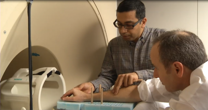
According to Foster, current imaging methods look at each bone individually and measure each angle, which does not capture how the bones move together. In order to overcome this problem, the team developed an approach called principal component analysis and imaged the wrist using an MRI scanner in various positions in radial-ulnar deviation. From these images, the bone surfaces were extracted and implemented with their statistical model to model how the bones move. Chaudhari stated that during the research process, the team had to generate their own data sets, such as MRI acquisition protocols, since movement is usually discouraged within MRI scanners due to the blurring of images.
“That’s part of the reason why I said we needed to collect our own data sets,” Chaudhari said. “[It’s] because it’s exactly the opposite of what is typically done. Technology says ‘don’t move’ and then you capture the image. Here we are actually telling people to move in the scanner.”
From the models they developed, the results affirmed that the left and right wrist move in a similar manner. Chaudhari explained how this information could be helpful for a surgeon doing reconstructive surgery.
“Previously, the physicians assumed that the right and left wrists move the same,” Foster said. “I think now we have more evidence that they do move the same, so you could use, if you have a wrist injury, you could use the uninjured hand to do surgical planning on the injured wrist.”
His team also discovered that when performing the maneuver of moving the wrist from left to right, men move in plane whereas women move a little out of plane with the wrist moving in a slight up and down motion. Foster stated that a possible explanation for this difference is that women have more laxity, or flexibility, whereas men have less range of motion which could influence the way the bones move. Chaudhari stated that this knowledge is important in developing treatment methods for men and women, since currently the same methods and techniques are used for both genders. With this new information, doctors could change the procedures according to how the wrist moves.
“So broadly in medicine, what we try to do is what we call precision medicine,” Chaudhari said. “This is essentially trying to tailor the treatment to an individual, so I think with methods of this nature, we are personalizing medicine to that individual because now we have the ability of knowing how each person’s individual wrist moves per say or track movements of their wrist and that is useful from the point of view of personalizing treatments.”
Anand Joshi, research assistant professor at University of Southern California, explained that their developed methodology will allow researchers to track and analyze bone changes during treatments.
“Improved tracking of bone changes in response to therapies will lead to reduced disease burden, toxicity, side-effects, and overall cost,” Joshi said. “Our methods have the potential to improve sensitivity to small, early changes in bone erosion status. The image analysis studies have the potential to provide new imaging biomarkers to be used in clinical trials evaluating the efficacy of new arthritis drugs.”
Chaudhari also said the technique will allow doctors and researchers to precisely identify problems in the wrist and learn more about how the wrist works.
“I think it’s not until recently that we have had the ability of taking in vivo imaging like MRI or CT fast and actually knowing exactly what’s happening inside the body,” Chaudhari said. “I think these techniques now are just taking you to the next level.”
Written by: Michelle Wong – science@theaggie.org


