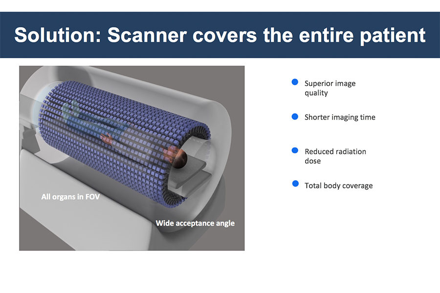
New device will be up to 40x more sensitive than standard scanners
A team of researchers at the UC Davis EXPLORER project are closing in on a goal over a decade in the making — the first PET scanner in the world that can scan the entire body at once rather than in 15 centimeter segments.
Simon Cherry, a distinguished professor of biomedical engineering, and Ramsey Badawi, an associate professor of radiology, first chatted about a possible total-body PET device in the mid-2000s. At the time, many of their colleagues in radiology and nuclear medicine considered such a project too unwieldy or expensive to be worthwhile. But grants from the National Cancer Institute’s Provocative Questions project and the UC Davis Research Investments in the Sciences & Engineering program allowed the team to produce promising prototype design plans. The National Institutes of Health Transformative Research Award — $15 million over five years — finally allowed the team to produce total-body PET scanners in two sizes.
“We’ve now built two prototypes — small-scale versions for scanning animals,” Badawi said. “The full human scanner should be finished in May of this year if all goes well.”
Positron emission tomography, also known as PET, is a sensitive medical imaging technique used in research and clinical settings. One of the most common uses for PET imaging today is for cancer diagnostics, although the team hopes to utilize the device in the future to examine systemic processes happening throughout the body in multiple organ systems.
“PET works by injecting a very small amount of a radioactively labeled compound into the body,” Cherry said. “For cancer imaging, the compound we use is an analogue of glucose. Glucose is a sugar, it’s used by the body to generate ATP as an energy source. Tumor cells are rapidly dividing, so they have high energy demands, and they’ll take up a lot of this radioactively labeled sugar. That’s how we get the contrast in our images where we can see these tumors very well.”
The total-body scanner will include all of the patient within its sensitive detectors, which collect energy emitted from the small doses of radiation injected into the patient.
“Most of the isotopes we use have a half-life between one to two minutes up to a few hours,” said Dr. Martin Judenhofer, an associate project scientist and an EXPLORER project manager. “FDG happens to have a half-life of two hours. After about ten half-lives, most of it is decayed.”
One benefit of the full-body scanner is the ability to use smaller amounts of radioactive tracers, such as fludeoxyglucose (FDG), to obtain quality images. FDG is a glucose compound tagged with radioactive fluorine. Lower doses of radionuclides will allow children and pregnant women to more safely use PET scanners for medical imaging.
“One of the interesting things we’ll be able to do is to look at signaling between different organs, which is not something we’ve been easily able to do before,” Badawi said.
Collecting much more of the emitted radiation will allow researchers and clinicians to generate images nearly 40 times more sensitive. A newfound flexibility in their procedures will follow.
“We have a choice,” Cherry said. “We can either get the image much more quickly — a 20-minute scan could be done in 30 seconds — or we can still scan for 20 minutes and get much higher-quality data.”
The MINI EXPLORER installed at the National California Primate Research Center has all of the components of the full-size scanner in a smaller package and has been used for research on animals including rhesus macaque monkeys. A second MINI scanner intended for the UC Davis veterinary school to be used with companion animals will be delivered in February 2018. The human scanner should arrive at the UC Davis Medical Center in Sacramento by 2019 and will be the crowning achievement for the EXPLORER team.
Written by: George Ugartemendia — science@theaggie.org
Editor’s note: An earlier version of this story stated that the human scanner would be finished in May of next year. It will be finished in May of this year. The article has been updated to reflect this change.


