The George Lab aims to model tumor microenvironments and physiological networks for cancer patients three dimensionally.
Though two-dimensional animal modeling has provided valuable information for human cellular interactions and disease mechanisms, it faces limitations. Human cells adopt different behaviors across two-dimensional and three-dimensional models.
In the human body, cells are surrounded by other cells from all sides in a dynamic environment. These interactions are difficult to capture in a two-dimensional cell model, such as cell culture, which produces flat layers of human cells in media. In animal modeling, promising cancer and cardiovascular therapy treatments identified in mouse models have led to toxic effects in humans. Consequently, the George Lab, part of the UC Davis biomedical engineering department, uses “organ-on-a-chip” technology to model complex cellular interactions and behaviors across human tissue and organ systems with cancer.
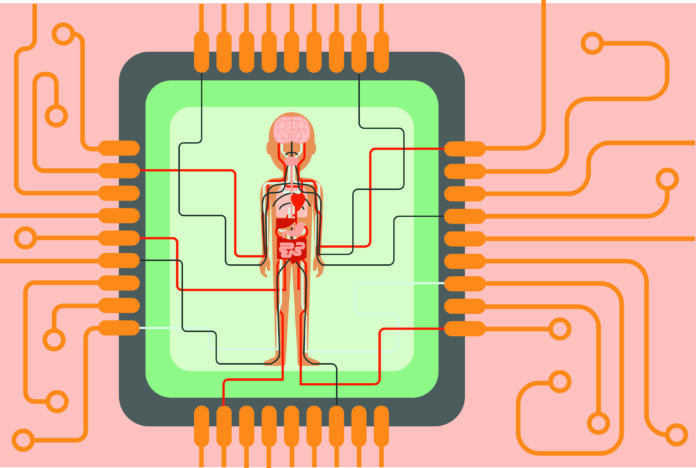
Organ-on-a-chip technology models intricate human organs inside a palm-sized, clear and flexible chip, complete with a vascular network and cell culture media to nourish cells three-dimensionally.
“It’s the same process of design that you would see on a circuit in a computer chip, except in this case, it’s microfluidics,” said Steven George, a professor in the UC Davis Department of Biomedical Engineering. “You’re actually passing fluids, cells and other components into these chips.”
The George Lab seeks to model breast cancer among physiological networks inside these chips.
Breast cancer is the second most frequent cancer diagnosis for women in the United States. Additionally, it is the second leading cause of cancer death.
“20 percent of women diagnosed with early stage breast cancer will develop the recurrent disease,” George said. “Breast cancer recurrence drops the five-year survival rates among patients from over 80 to 20 percent.”
The George Lab aims to understand breast cancer dormancy and recurrence, along with the early steps in cancer metastasis that allows these processes to occur. In turn, organ-on-a-chip technology can be used as a clinical tool to develop personalized medicine. For breast cancer patients, chips could be generated to grow samples of tumors, administer chemotherapy trials and determine optimal chemotherapy strategies for each patient.
“Breast cancer tumors manipulate the bone marrow environment,” said Drew Glaser, a postdoctoral scholar in the George Lab. “The bone marrow is a space for white blood cells that work to remove cancers.”
Breast cancer recruits normal immunosuppressive cells and redefines the bone marrow environments.
“If you think about bone marrow, most of the time you think about the soft, squishy tissue,” Glaser said. “There is also hard pieces of bone interspersed among the soft, squishy tissue.”
Breast cancer can metastasize in either of these bone marrow microenvironments. Bones constantly and gradually remodel their environments, maintaining a balance between bone growth and clearance. Breast cancer tumors disrupt this balance, preferring active osteoclast cells, bone-clearing cells, over osteoblasts, bone-forming cells. By preferring bone clearance, the remodeled bone environment leads to bone destruction and creates a challenge for cancer treatment.
“The remodeled bone environment becomes difficult to treat,” Glaser said. “These cancers become chemorefractive, meaning they do not respond to chemotherapy. Patients can be exposed to radiation to remove the tumor, but this is when patients are at risk for developing fractures and pain, which can lead to morbid secondary outcomes.”
Glaser investigated the morphology and vessel network interactions between the two bone marrow environments through cancer migration assays, which involves physically separating the two environments inside of the chip while maintaining a small connection that links them. Glaser hopes to determine how each bone marrow environment shapes breast cancer metastasis and tumor aggression.
“Our current goal is to fabricate a representative bone marrow model of healthy patients, then we can see how cancer cells navigate in this environment,” said Aravind Anand, a fourth-year biomedical engineering major.
Organ-on-a-chip technology shows promise for mapping complex relationships between human organ environments and breast cancer cells, which could eventually lead to more ideal medical treatment for patients.
“While bone metastasis is the intended application of this project, this device may ultimately be able to model many other diseases or provide a platform to study hematopoiesis,” Anand said.
Written by: Foxy Robinson – science@theaggie.org


